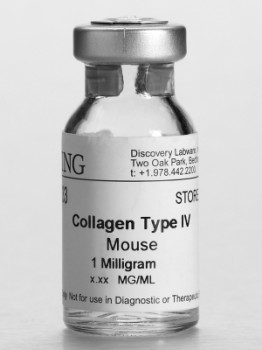
Corning® Collagen
By Becton Dickinson
Corning extracellular matrices (ECMs) enable researchers to mimic in vivo environments for 2D and 3D cell culture applications.
Corning offers a broad range of collagen types that are derived from multiple species. These collagens have been used to culture a variety of cell types to promote adhesion, growth, and/or differentiation.
- Collagen I is found in most tissues and organs, but is most plentiful in dermis, tendon, and bone. It can be used as a thin layer on tissue-culture surfaces to enhance cell attachment and proliferation, or as a gel to promote expression of cell-specific morphology and function. Type 1 collagen is commonly used to culture endothelial cells, hepatocytes, muscle cells, and a variety of other cell types.
- Type II Collagen is the pricipal collagenous component of cartilage, intervertebral disc and vitreous humour. It is also found in other tissues during development. Its main function is to provide tensile strength and give cartilage the ability to resist shearing forces. Type II Collagen supports chondrocyte adhesion and may influence the differentiated phenotype of these cells. It can also be used as an in vivo model in rats and mice for arthritis studies (injection of Bovine Collagen II induces arthritis).
- Type III Collagen is found co-localized with Type I Collagen in tissues, blood vessels and skin. Type III Collagen is a homotrimeric procollagen comprised of three identical pro a, (III) chains. Collagen III is important for the development of skin and the cardiovascular system and for maintaining normal physiological functions in adult life. Collagen III has been used for studies of Collagen I fibrillogenesis in normal cardiovascular development. Many of the same properties and functions of animal derived collagens can be duplicated using recombinant human collagens. FIBROGEN Recombinant Human Collagens Type I and III are highly purified, reliable and consistent. They provide alternatives to animal derived collagens for biomedical applications in which an enhanced safety profile, and greater reproducibility and quality are critical.
- Collagen VI is a large, multidomain extracellular matrix protein. Its heterotrimeric chains assemble into a microfibrillar network via tetramerization and end-to-end association1. It‘s pattern of distribution and its unique structure and expression compared with other ECM molecules indicate that Collagen VI may fulfill specialized tasks in tissue organization and cell functioning.
- Collagen V is found in whole placenta, amnion, chorion, and cornea. It can be used as a thin coating on tissue-culture surfaces to study collagen V effects on cell behavior. Collagen V has been shown to inhibit endothelial cell proliferation selectively.
- Collagen VI is a large, multidomain extracellular matrix protein. Its heterotrimeric chains assemble into a microfibrillar network via tetramerization and end-to-end association1. It‘s pattern of distribution and its unique structure and expression compared with other ECM molecules indicate that Collagen VI may fulfill specialized tasks in tissue organization and cell functioning.
