Albany Advanced Digital Imaging - Whole Slide Imaging Services
By Albany Advanced Digital Imaging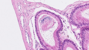
An affordable solution for your microscopy needs!
Albany Advanced Digital Imaging offers whole slide imaging by utilizing cutting edge technology to produce detailed digital scans of an entire slide - in minutes - to create a single-stitched high resolution image file, with a magnification range of 0.5x-80x. Services by Albany Advanced Digital Imaging will accelerate your research projects and digital archival processes by greatly reducing the time and effort required by traditional microscopy methods.
Brightfield imaging with 1"x3" standard slide format is currently available, with optional Z-stack functions. Your data is accessible via secure cloud storage or physical storage device. Fluorescence-capabilities will be added soon!
Order fulfillment process: Follow instructions to submit your order with Krackeler. Once submitted, Krackeler will forward your details to Albany Advanced Digital Imaging. A rep from AADI will then reach out to you with all pertinent logistical and lead time information.
| Cells & Tissue Stains | |
| Bismarck Brown Y | Stains mucins yellow to light brown |
| Carmine | Used to stain nuclei, glycogen, and acid mucopolysaccharides red or crimson in color |
| Congo Red | Stains amyloid deposits in animals and the cell walls of plants and fungi in color from pink to dark red |
| DAB | HRP substrate for single antigen detection antigen = brown |
| H&E | Hematoxylin, nuclei = purplish blue; Eosin, cytoplasm, and extracellular matrix = pink |
| Lugol's Iodine | Indicates the presence of starches in plants and animals by turning dark blue to black. It is also useful in identifying cancerous and precancerous lesions - healthy cells contain glycogen and are stained brown, while the cancer cells remain unstained |
| Methyl Green | Stains nuclei green, alternative to Hematoxylin depending on other substrates used |
| Methylene Blue | Detects DNA and RNA in the nucleus and cytoplasm, turning blue. Also used for bacterial identification as well as cell structure in both plant and animal cells |
| Mucicarmine | Used to detect mucin, identifying microorganisms and some spores and fungi, color ranges from light pink to red |
| NovaRED | HRP substrate for single antigen detection, antigen = red |
| Nuclear Fast Red (Kernechtrot Red) | Nuclear chromatin = red |
| Oil Red O | Stains fat, lipids, and triglycerides red |
| Orcein | A reddish-brown stain that is used to visualize chromosomes and elastic fibers in tissue sections |
| PAS - Periodic Acid-Schiff Reaction | Stains polysaccharides/glycoproteins/glycolipids/mucin red to purple |
| Prussian Blue | Detects the presence of iron in cells and tissue, producing a blue stain |
| PSH - Picrosirius Red & Hematoxylin | Nuclei = purple, collagen = red, cytoplasm = pink to light violet |
| Safranin | Stains nuclei red. It can also be used to stain mast cells, mucin, and cartilage |
| Silver Stain | Used to detect proteins and nucleic acid resulting in a dark brown to black stain |
| Sudan Black | Stains lipids/triglycerides/fatty acids as well as neutrophils in blood and bone marrow black |
| Toluidine Blue | Used to stain nuclei = blue, mast cells = purple, cartilage = purple, mucins = red to purple |
| Trichrome Stains | Gomori: nuclei = black, collagen = blue to green, cytoplasm and muscles = red |
| Lillie's: nuclei = black, cytoplasm = brown, muscle = red, collagen = blue to green | |
| Mallory's: nuclei and muscles = pink to red, collagen = blue, erythrocytes = orange | |
| Masson's: nuclei = dark brown to black, collagen and bone = blue to green, cytoplasm/keratin/muscles = red to pink | |
| Wright-Giemsa Stain - Differentiates between blood cell types, and used in blood smears, bone marrow, urine samples and parasites/bacteria, nuclei = blue to purple, cytoplasm = pink to orange | |
| Bacterial, Yeast, and Fungi Staining | |
| Acid Fuchsin | Stains growing bacteria as well as muscle tissue red |
| AFS - Acid-Fast Stain | Bacterial and fungal detection, positive acid-fast bacteria = red, negative = blue to green |
| Basic Fuchsin | Stains bacteria and nuclei red |
| Crystal Violet | stains nuclei, proteins, and Gram-positive bacteria purple |
| Gram Stain | Used to differentiate between gram-positive and gram-negative bacteria, positive = purple, negative = pink |
| Malachite Green | Detects endospores in bacteria, results in a green coloration |